surgical data
socket lifting
| patient information | |
|---|---|
| age | 61 |
| gender | Old Male |
| implant |
#15, 16, 17, 27 SST Implants |
socket lifting
Internal Hex Fixture : SST Type
Fig 01
Implant placement combined socket lifting
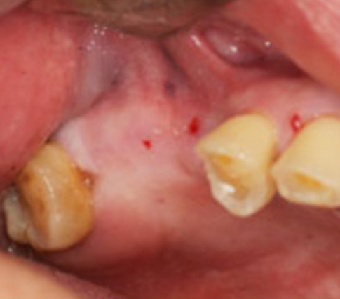
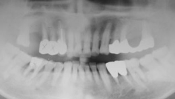
Fig 02
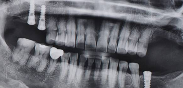
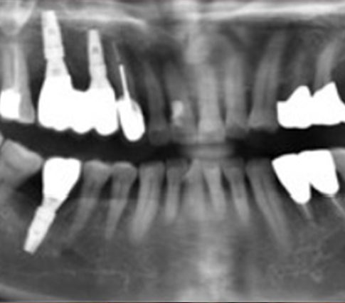
Initial panorama x-ray
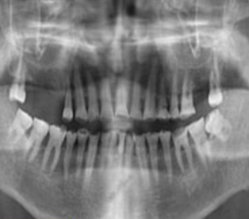
Fig 03
Sinus grafting performed by Crestal approach
on the #16,17 area
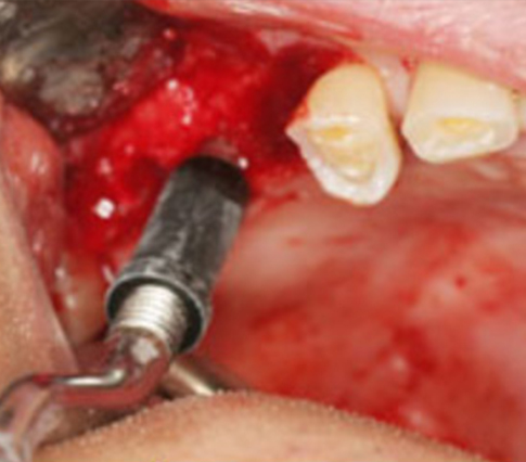
Fig 04
SG Implants were installed. Pictures of SST system
#15,16 SFSR411 / #17 SFSW511
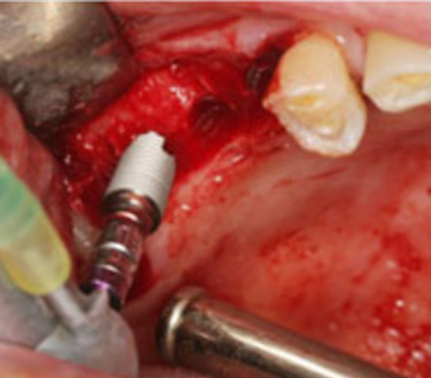
Fig 05
Primary stability checked with Torque Wrench
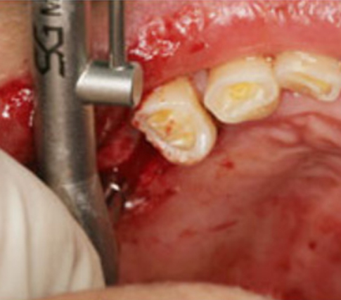
Fig 06
The picture taken after the SST implants placed
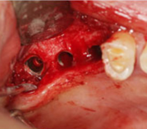
Fig 07
Healing abutments were inserted
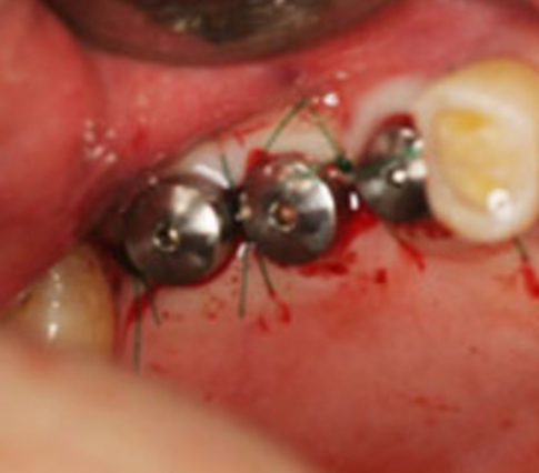
Fig 08
#14, 15, 16 SST implants installed & Healing abutments were inserted. / #27 implant was inserted combined socket lifting
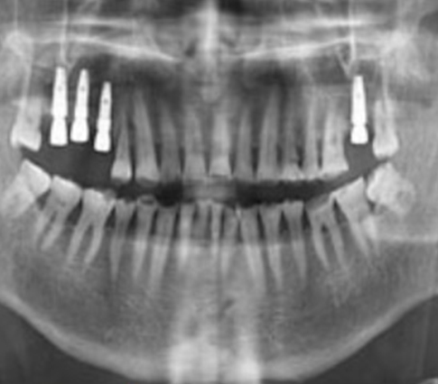
Fig 09
Healing sequences of sinus area
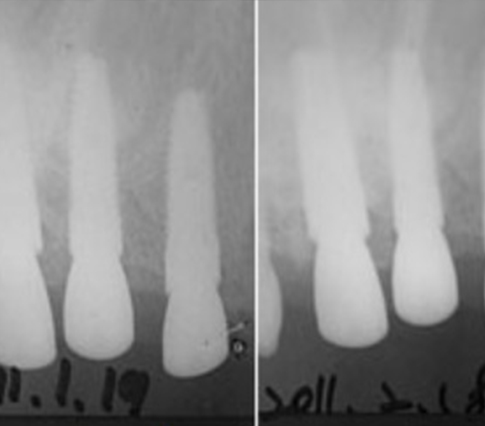
Fig 10
Impression taking with transfer type
on the fixture level
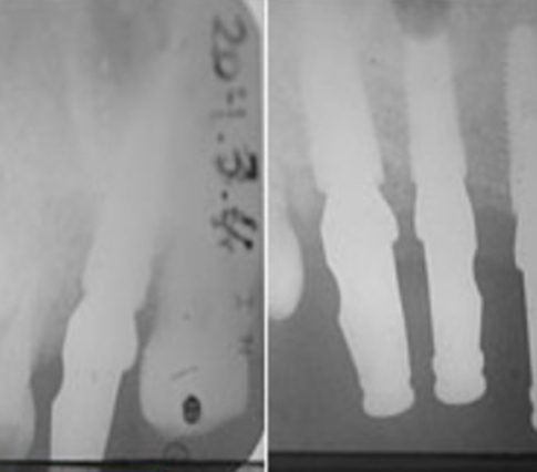
Fig 11
G-mask state, trimming was done
for gingival forming
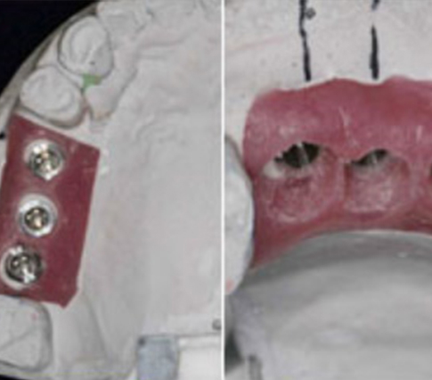
Fig 12
G-mask state, trimming was done
for gingival forming
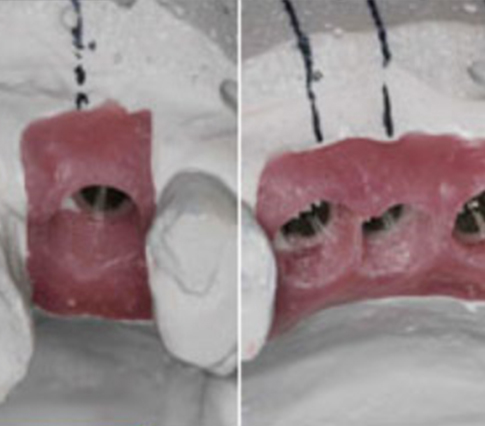
Fig 13
Abutment try-in
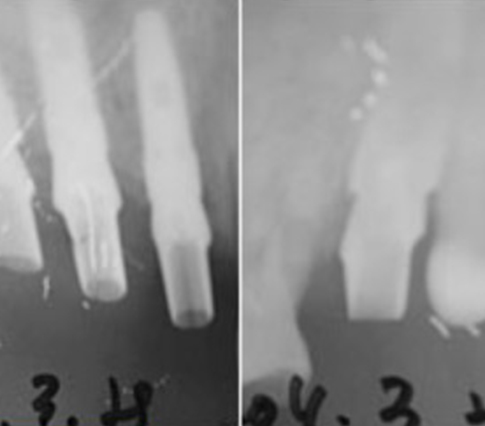
Fig 14
Resin cap try-in
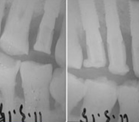
Fig 15
Final prosthesis
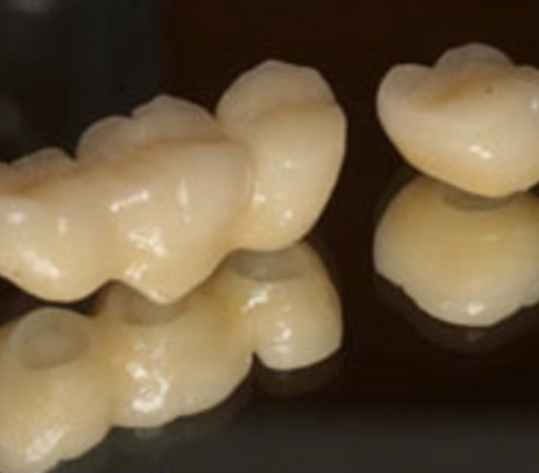
Fig 16
The pictures taken after the final prosthesis
on the Rt Molar
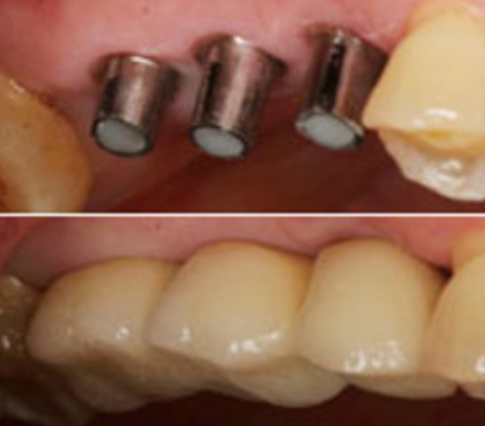
Fig 17
The pictures taken after the final prosthesis
on the LT 2nd molar after 5 months
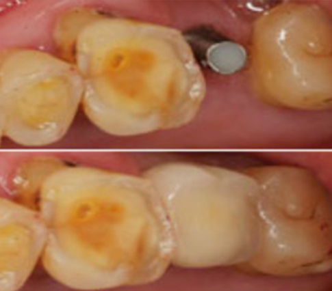
Fig 18
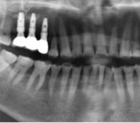
Final prosthesis x-rays
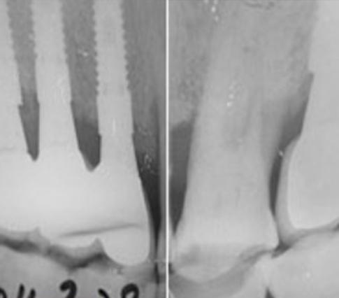
Fig 19
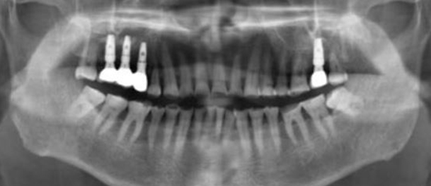
Finished panoramic x-ray
surgical data
Combined Gbr
| patient information | |
|---|---|
| age | 59 |
| gender | Old Male |
| implant |
#14, 16 : GBR → SST implants placement #46 : SST implants placement #26, 27 : sinus graft + simulaneous SST implants placement #36 : immediate SST implants placement + GBR |
combined gbr
Internal Hex Fixture : SST Type
Fig 01
#14, 15 teeth extraction and sinus grafting performed
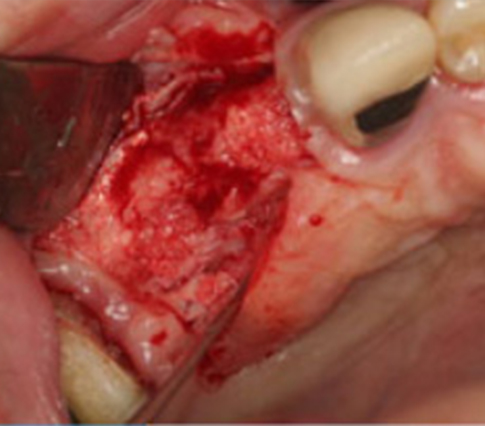
Fig 02
Tension free suture was done through undermining after GBR.
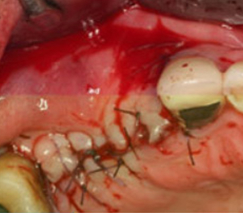
Fig 03
5 months later the GBR
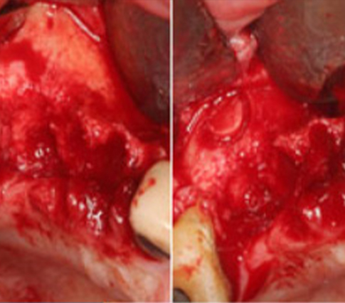
Fig 04
Using the guide-pins for path & position of the implants
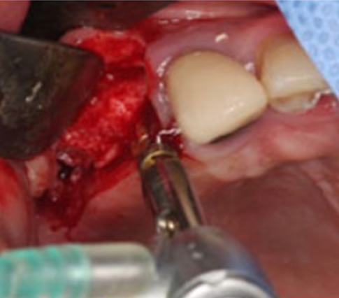
Fig 05
Drilling the bone
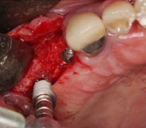
Fig 06
SG Implants were installed.
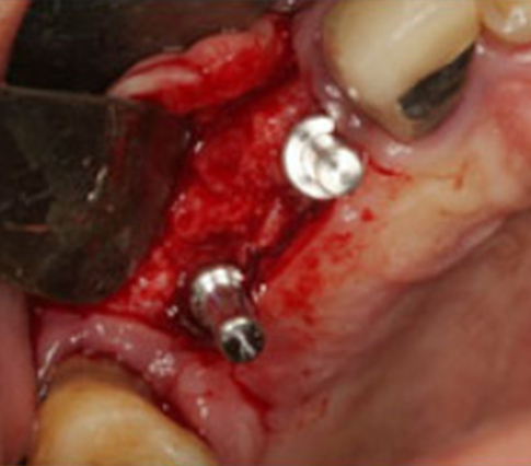
Fig 07
The picture taken after the SST implant placed
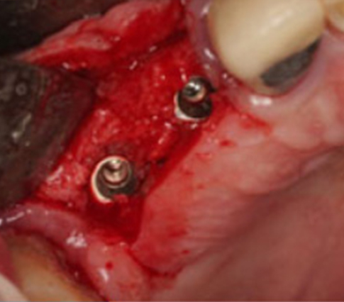
Fig 08
Healing abutments were inserted
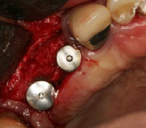
Fig 09
Palacci flap was done for soft tissue management
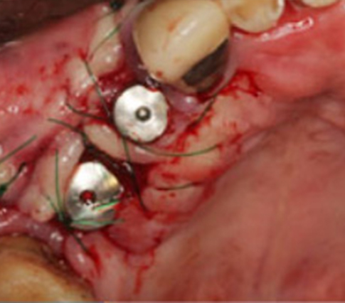
Fig 10
Mandible right molar area was scheduled for implant
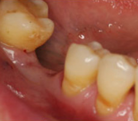
Fig 11
There was a sufficient bony volume
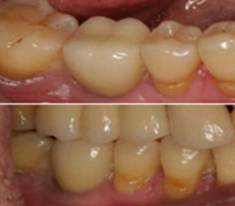
Fig 12
1-stage implant was performed because of good primary stability
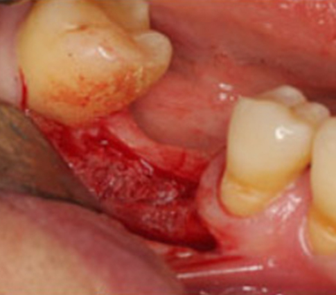
Fig 13
Torque Wrench
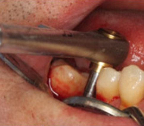
Fig 14
The pictures taken after the prosthesis treatment completed after 3 months implant placement
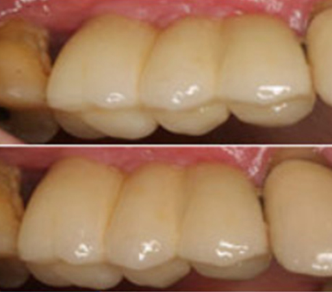
Fig 15
The picture taken after the prosthesis treatment completed after 3months later dental implant placement 2nd molar will be extracted
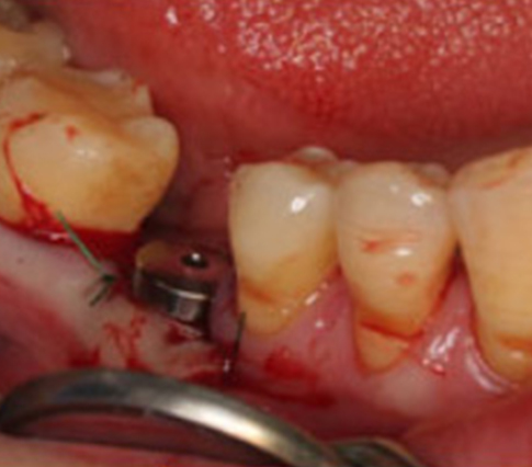
Fig 16
Panoramic picture taken after the prosthesis treatment
surgical data
Flapless Surgery
Way to place Dental Fixture without incision of the gums
Reduce bleeding, pain, and swelling to a minimum.
Reduced number of complaints due to shorter treatment period and recovery period.
| patient information | |
|---|---|
| age | 43 |
| gender | Female |
| implant |
#16, 17, 36 IT implants |